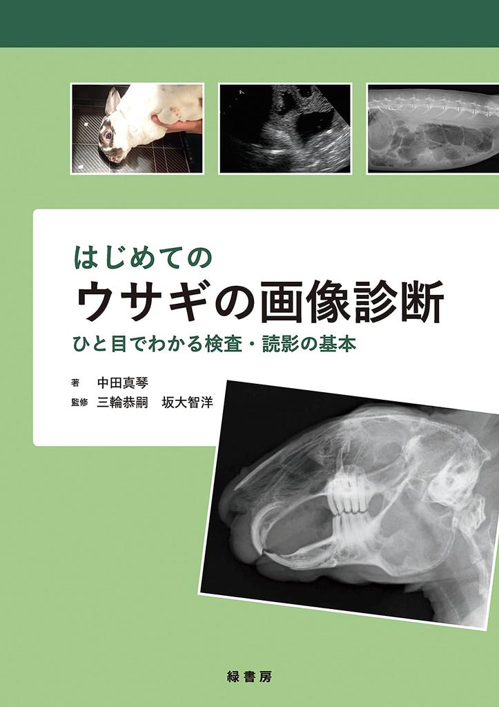
First time imaging diagnostics for rabbits: the basics of examination and interpretation at a glance


 Reembolso por falta de entregaGarantía de calidadLogística seguraProtección de la privacidad
Descripción
The only diagnostic imaging book specifically for rabbits!
This book provides easy-to-understand explanations of rabbit diagnostic imaging basics and testing know-how essential for clinical veterinarians, with over 700 images and illustrations. This book is useful for everyone, from those new to rabbits to those looking to refresh their existing methods.
[Key Features of This Book] ● This popular series from the veterinary journal "Companion Animal Diagnostic Imaging" has been significantly expanded and revised into a book. With numerous new illustrations and photographs, it's approximately 100 pages longer than the original series. ● Basic anatomy and physiology are explained in an easy-to-understand manner, including differences between dogs and cats. ● Detailed explanations of what to look for when assessing normal and abnormal images. ● Basic information on pathology and treatment methods are provided for each disease. ● Carefully explained are essential techniques for everyday examinations, such as safe restraint methods, X-ray techniques, and ultrasound probe movements.
Table of Contents Appendix X-ray Images of a Normal Rabbit
Chapter 1: Basics of Imaging in Rabbits Introduction: Before Beginning Imaging 1. X-ray Examination: Imaging Methods Examination Preparation and Imaging Conditions Cephalic X-rays Chest and Abdominal X-rays Extremity X-rays When to Use X-rays Abdominal Contrast X-rays Column: Sedatives Used in Rabbits 2. Ultrasound Examination: Scanning Methods Examination Preparation Restraint Methods Screening Scan Procedure and Precautions When to Use Ultrasound 3. Practical CT and MRI Examinations CT Examination MRI Examination
Chapter 2: Head 1. Dental Disease Introduction Anatomy and Physiology Normal Images Malocclusion Dental-Related Diseases Point: Key Points in Imaging Examinations 2. Head Fractures and Tumors Introduction Anatomy and Normal Images Maxillary Fractures Mandibular Fractures Neoplastic Diseases of the Head (Intraoral) Other Non-Dental Diseases of the Stomatognathic System Point: Key Points in Imaging Examinations 3 Ear Canal Diseases Introduction Anatomy and Normal Images Abscesses at the Base of the Ear and Tympanic Bulla Point: Key Points in Imaging Examinations 4 Nasal and Sinus Diseases Introduction Anatomy, Physiology, and Normal Images When to Use Imaging Examinations Rhinitis and Sinusitis Abscess Nasal Passage Obstruction Due to Dental Disease Tumor Foreign Body Point: Key Points in Imaging Examinations
Chapter 3 Chest 1 Lung, Anterior Mediastinal, and Esophageal Diseases Introduction Anatomy and Normal Images When to Use Imaging Examinations Pneumonia, Tracheal and Bronchitis, Lung Abscess Lung Tumor Precordial Mass (Thymoma, Lymphoma) Diaphragmatic Hernia Megaesophagus Point: Key Points in Imaging Examinations 2 Cardiovascular Diseases Introduction Anatomy and Physiology Normal Images and Evaluation Methods Congestive Heart Failure Pericardial Effusion Cardiomyopathy Congenital Heart Disease Arteriosclerosis Point: Key Points in Imaging Examinations
Chapter 4 Abdomen 1 Gastrointestinal Diseases, Intra-Abdominal Abscesses Anatomy and Normal Images When to Use Imaging Tests Indigestion Gastrointestinal Obstruction by Fecal Mass Acute Gastric Dilatation Intra-Abdominal Abscess Point: Key Points in Imaging Tests
2 Liver Diseases Introduction Anatomy and Physiology Normal Images Gastrosteroid-Induced Hepatitis and Hepatic Lipidosis Bacterial Hepatitis Hepatic Lobe Torsion Liver Tumors Point: Key Points in Imaging Tests
3 Urinary Diseases Introduction Anatomy, Physiology, and Normal Images Urolithiasis Renal Failure Renal Cysts Urinary Neoplastic Diseases Point: Key Points in Imaging Tests
4 Reproductive Diseases Introduction Anatomy and Physiology Normal Images Uterine Diseases Ovarian Diseases Pregnancy Diagnosis Testicular Diseases Point: Key Points in Imaging Tests
Chapter 5 Skeleton 1 Bone and Orthopedic Disorders Introduction Anatomy and Normal Images Fractures Tumor Spondylosis, Osteoarthritis/Congenital Spinal Deformity Splayfoot Sore Hock, Osteolysis Due to Abscess Traumatic Joint Dislocation/Subluxation Points to Note in Imaging Examinations